
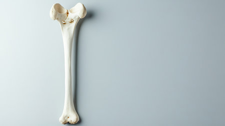
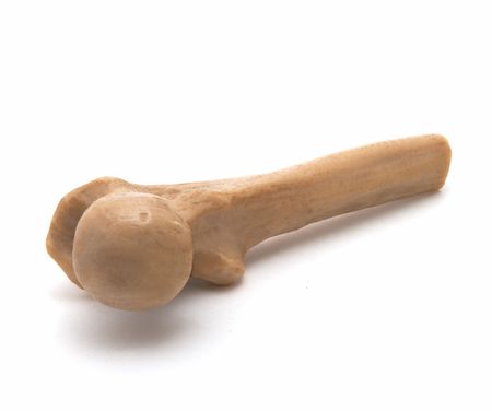
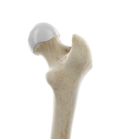
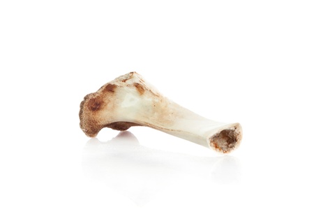
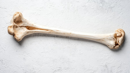
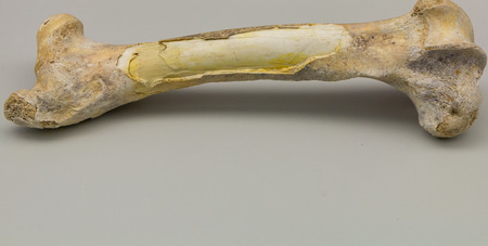
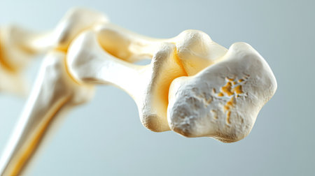
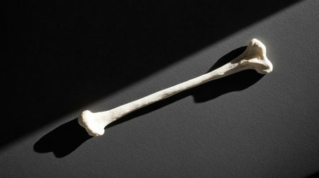
A detailed view of a human femur bone displayed against a simple background. Ideal for medical, educational, and anatomical projects focused on the human skeleton.
Коллекция по умолчанию
Коллекция по умолчанию
Создать новую
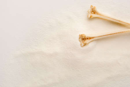
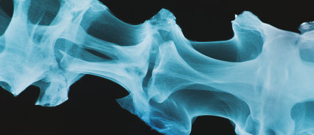
White Collagen powder background with bones. Natural beauty and health supplement for skin and bones.
Коллекция по умолчанию
Коллекция по умолчанию
Создать новую

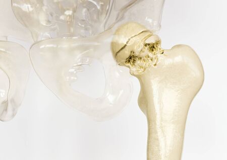
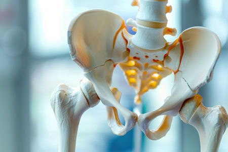
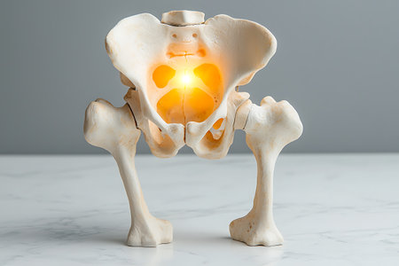
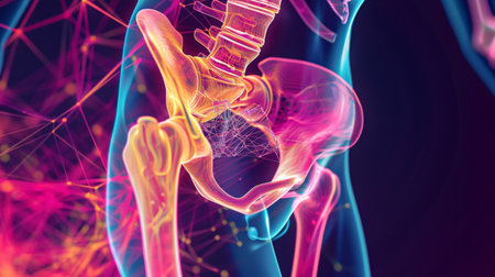
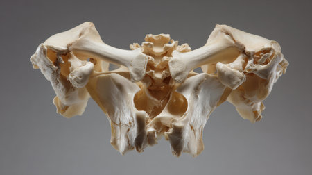
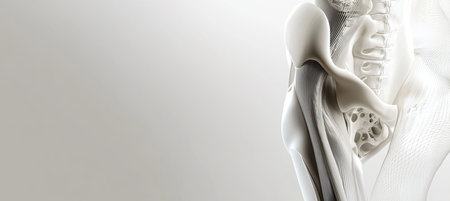
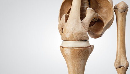
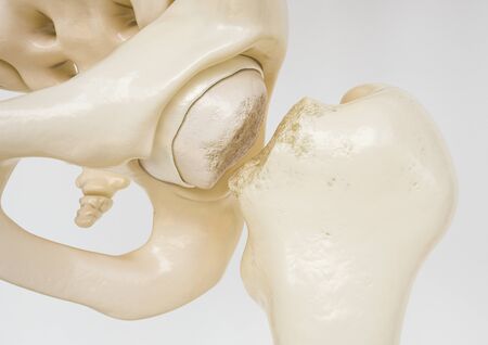
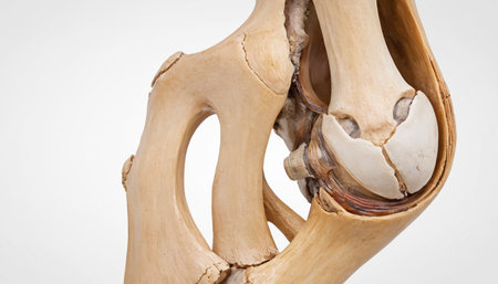
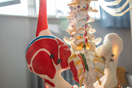
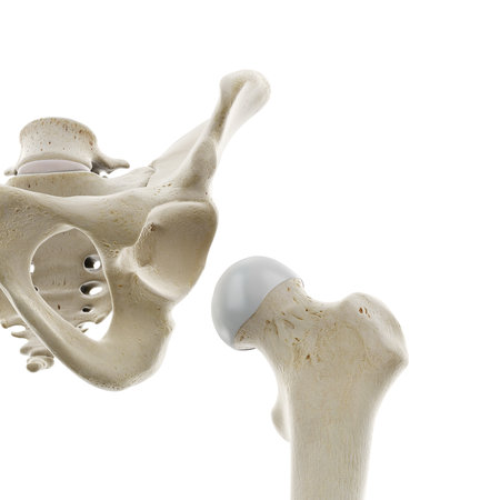
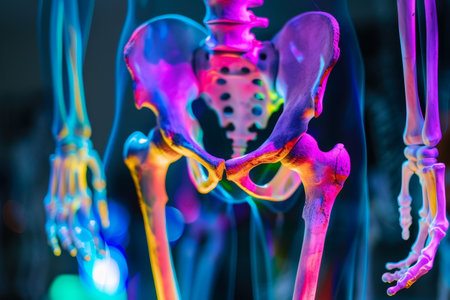
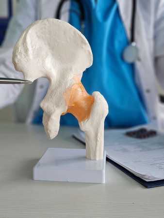
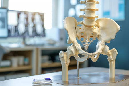
A close-up view of a human hip joint anatomical model, highlighting the intricate structure of the bones and ligaments. The model is set against a light blue background, emphasizing the detail and complexity of the joint.
Коллекция по умолчанию
Коллекция по умолчанию
Создать новую
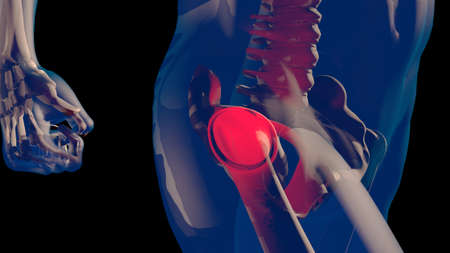
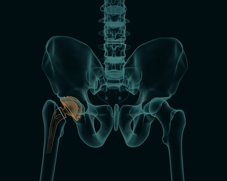
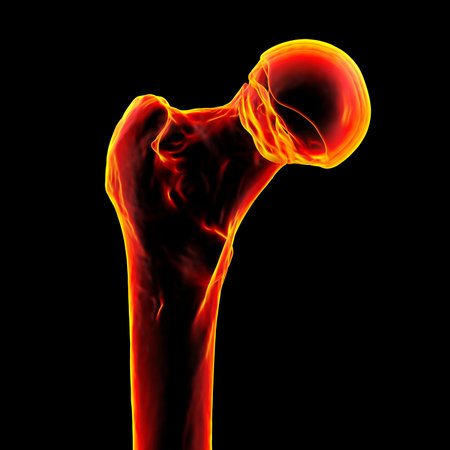
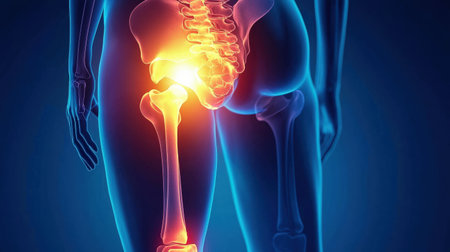
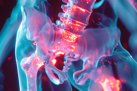
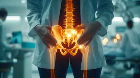
Thigh Femoral Head and Neck Pain in Human Body Transparent Design 3D Illustration
Коллекция по умолчанию
Коллекция по умолчанию
Создать новую
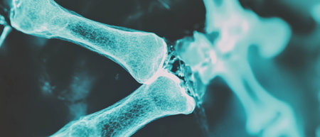
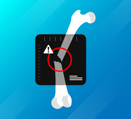
X-ray showing details of a broken bone with sharp fragments in medical diagnosis
Коллекция по умолчанию
Коллекция по умолчанию
Создать новую
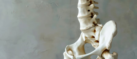
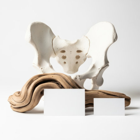
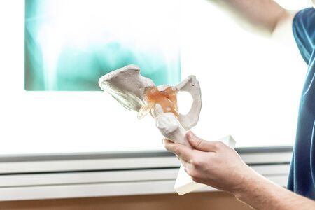
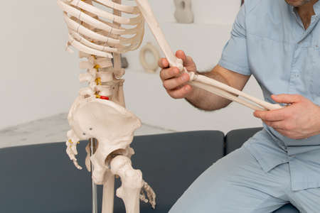
A white, plastic model of the human pelvis and spine stands against a grey wall. The model's bones are detailed, and the curved spine is positioned vertically, with the pelvis positioned below.
Коллекция по умолчанию
Коллекция по умолчанию
Создать новую
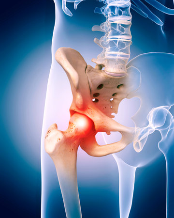
Severe osteoarthritis of the hip - 3D Rendering
Коллекция по умолчанию
Коллекция по умолчанию
Создать новую
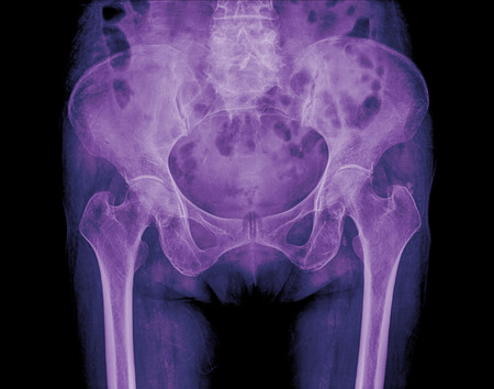
X-ray image of a abdomen, showing bones and internal organs on a veterinary clinic screen.
Коллекция по умолчанию
Коллекция по умолчанию
Создать новую
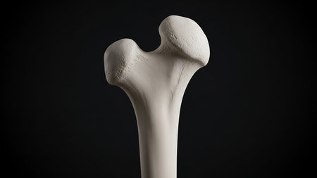
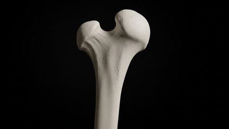
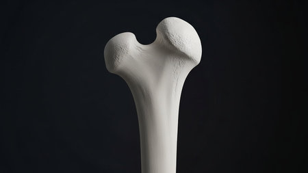
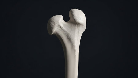
Detailed view of a human femur bone against a dark background, highlighting its anatomy.
Коллекция по умолчанию
Коллекция по умолчанию
Создать новую
osteoporosis thigh bones from the inside- 3d rendering
Коллекция по умолчанию
Коллекция по умолчанию
Создать новую

Isolated shin bone displayed on a plain white background, showcasing details of the skeletal structure. Ideal for medical, educational, and anatomical uses.
Коллекция по умолчанию
Коллекция по умолчанию
Создать новую
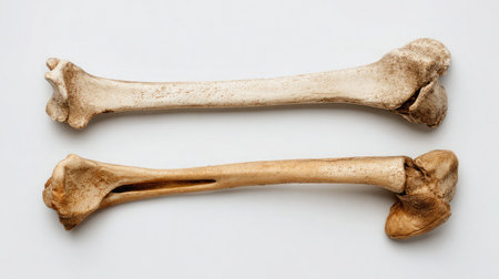
Top-down view of separated femur and tibia bones of the knee laid flat on white
Коллекция по умолчанию
Коллекция по умолчанию
Создать новую
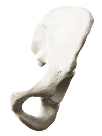
Pelvic bone model isolated on white background
Коллекция по умолчанию
Коллекция по умолчанию
Создать новую

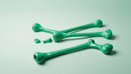
Green bones for make-up on a light background.
Коллекция по умолчанию
Коллекция по умолчанию
Создать новую
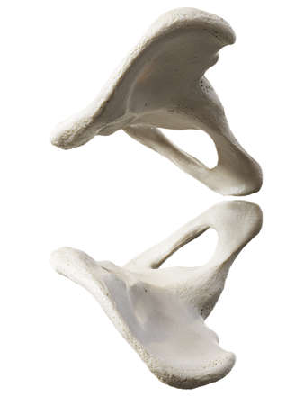
Two bones of the lower body, one of which is a hip, are shown in a close up. The bones are white and appear to be in a distorted or warped position. The image has a surreal or dreamlike quality to it
Коллекция по умолчанию
Коллекция по умолчанию
Создать новую

Medical poster image of the bones of the pelvis, the joint in the hip part. Arthritis, inflammation, fracture, cartilage, . Copy space, 3D illustration, 3D render
Коллекция по умолчанию
Коллекция по умолчанию
Создать новую
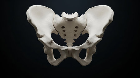
Detailed view of human pelvis bone anatomy isolated on black
Коллекция по умолчанию
Коллекция по умолчанию
Создать новую

bones and skull of an unknown animal in the Spitalfields Market
Коллекция по умолчанию
Коллекция по умолчанию
Создать новую
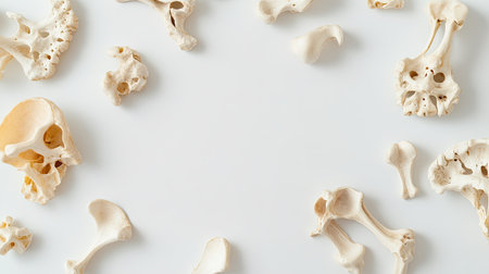
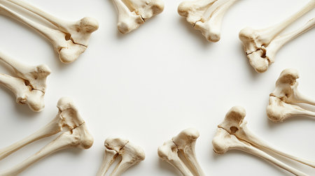
A collection of human bones arranged on a light background, suitable for educational, scientific, or artistic purposes. Showcases anatomy and structure.
Коллекция по умолчанию
Коллекция по умолчанию
Создать новую
Human skeleton with burning candle on white marble table. 3d rendering
Коллекция по умолчанию
Коллекция по умолчанию
Создать новую
Piece of a bone on a white background
Коллекция по умолчанию
Коллекция по умолчанию
Создать новую

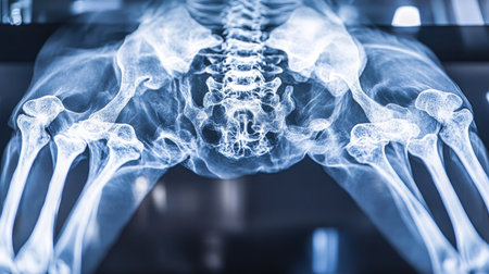
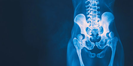
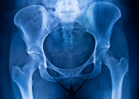
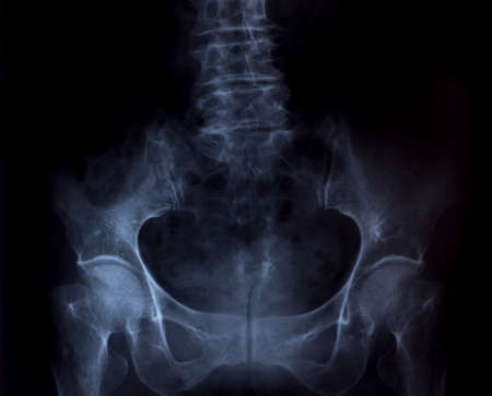
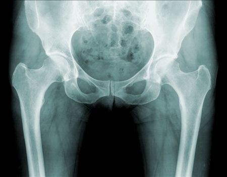
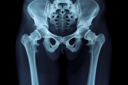
X-ray of pelvic bones
Коллекция по умолчанию
Коллекция по умолчанию
Создать новую
Lumbar Spine and Pelvis XRay Highlighting Skeletal Structure and Medical Imaging Techniques.
Коллекция по умолчанию
Коллекция по умолчанию
Создать новую
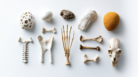
A collection of bones and other objects, including a skull, are arranged in a pattern on a white background. Concept of curiosity and fascination with the natural world
Коллекция по умолчанию
Коллекция по умолчанию
Создать новую
A digital illustration of the human hip joint, showcasing the ball-and-socket structure and range of motion in the hip region.
Коллекция по умолчанию
Коллекция по умолчанию
Создать новую
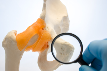
Doctor or medical student looks at pubic bone namely pubic symphysis through magnifying glass with selective focus on increase surface of pubic cartilage on anatomical models of pelvic bone
Коллекция по умолчанию
Коллекция по умолчанию
Создать новую
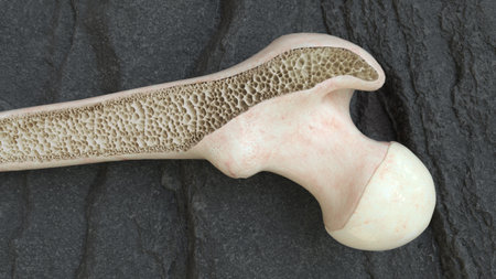
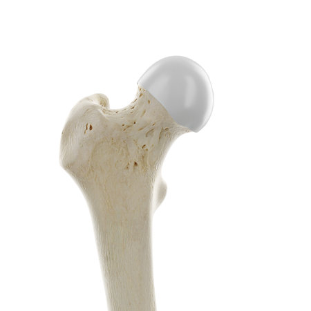
3d rendered medically accurate illustration of the upper femur joint
Коллекция по умолчанию
Коллекция по умолчанию
Создать новую
Anatomical representation of a human pelvis with clear details for educational use. Ideal for biology, anatomy studies, and medical illustrations in various fields.
Коллекция по умолчанию
Коллекция по умолчанию
Создать новую

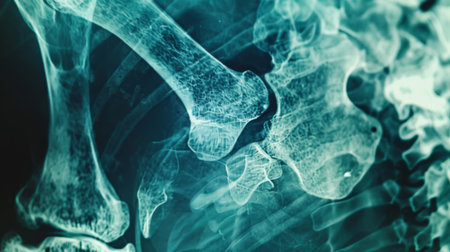
x-ray image of hip and show degenerative change or avascular necrosis hip
Коллекция по умолчанию
Коллекция по умолчанию
Создать новую
hip joint in artificial semi-anatomical model of pelvis and femur. Medical photos from consulting traumatologist or orthopedic surgeon.
Коллекция по умолчанию
Коллекция по умолчанию
Создать новую
x-ray of hip
Коллекция по умолчанию
Коллекция по умолчанию
Создать новую
tulips in basket isolated on white background. colors
Коллекция по умолчанию
Коллекция по умолчанию
Создать новую
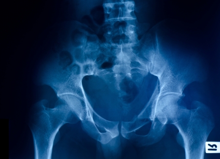
Hips pelvis xray scan test result.
Коллекция по умолчанию
Коллекция по умолчанию
Создать новую
Hip Endoprosthesis Display with Anatomical Chart for Medical Education and Training
Коллекция по умолчанию
Коллекция по умолчанию
Создать новую
3d rendered illustration of the hip bone
Коллекция по умолчанию
Коллекция по умолчанию
Создать новую
Realistic 3D Render of Human Skeleton anatomy - Back view
Коллекция по умолчанию
Коллекция по умолчанию
Создать новую
Detailed view of a human femur bone against a dark background
Коллекция по умолчанию
Коллекция по умолчанию
Создать новую

Close up of male doctors hand pointing at sacroiliac joint on skeleton spine model
Коллекция по умолчанию
Коллекция по умолчанию
Создать новую
Detailed image of a human femur bone isolated on a dark background, highlighting its anatomy.
Коллекция по умолчанию
Коллекция по умолчанию
Создать новую
Human skeleton with a metal hip prosthesis concept arthroplasty 3d render X-ray image
Коллекция по умолчанию
Коллекция по умолчанию
Создать новую
X-Ray Image Of Human Chest for a medical diagnosis
Коллекция по умолчанию
Коллекция по умолчанию
Создать новую
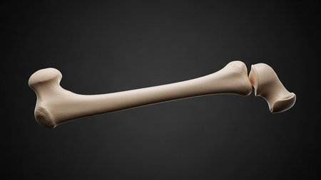
Femoral neck fracture - 3D Rendering
Коллекция по умолчанию
Коллекция по умолчанию
Создать новую
A single bone fragment placed diagonally on a textured white surface, showcasing its natural shape and detail in a clean, minimalistic style.
Коллекция по умолчанию
Коллекция по умолчанию
Создать новую
Human skeleton anatomy on white background. 3D illustration. Medical concept
Коллекция по умолчанию
Коллекция по умолчанию
Создать новую

Inflamed Human Knee Joint, 3D Illustration of Anatomical Structure, Highlighting Painful Condition
Коллекция по умолчанию
Коллекция по умолчанию
Создать новую
Thoracic spine with spinal, spine bones, disc and hip bone of human skeleton model for medical education
Коллекция по умолчанию
Коллекция по умолчанию
Создать новую
Green bones for make-up on a light background.
Коллекция по умолчанию
Коллекция по умолчанию
Создать новую
Detailed image of a white femur bone against a dark background, highlighting its anatomical structure
Коллекция по умолчанию
Коллекция по умолчанию
Создать новую

X-ray of the pelvis and spinal column
Коллекция по умолчанию
Коллекция по умолчанию
Создать новую
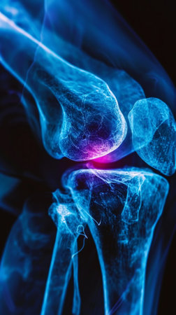
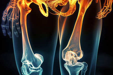
Blue x-ray photograph of knee pain, pain glows red. Trauma concept. Medical checkup. Generative AI.
Коллекция по умолчанию
Коллекция по умолчанию
Создать новую
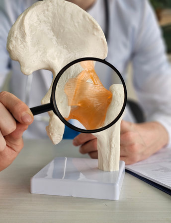
Medical professional examines hip joint model using magnifying glass during anatomy lesson
Коллекция по умолчанию
Коллекция по умолчанию
Создать новую
3d rendered medically accurate illustration of the hip joint
Коллекция по умолчанию
Коллекция по умолчанию
Создать новую
Subcapital femur fracture, 3D illustration showing a break just below the femoral head, near the hip joint.
Коллекция по умолчанию
Коллекция по умолчанию
Создать новую
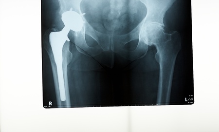
A x-ray of a hip prosthesis replacement
Коллекция по умолчанию
Коллекция по умолчанию
Создать новую

Physician, intern or student of medical university holds in his hand MRI (CT) scans of hip joint, comparing it with anatomical model of hip joint, located little further (focusing on model)
Коллекция по умолчанию
Коллекция по умолчанию
Создать новую
This image displays crossed femur bones arranged elegantly on a smooth white background. Ideal for medical illustrations, educational content, and anatomical studies.
Коллекция по умолчанию
Коллекция по умолчанию
Создать новую
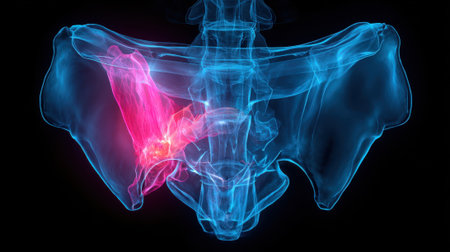
X-ray of a pelvis showing sacroiliac joint inflammation
Коллекция по умолчанию
Коллекция по умолчанию
Создать новую

Close up bone x-ray medical science background
Коллекция по умолчанию
Коллекция по умолчанию
Создать новую
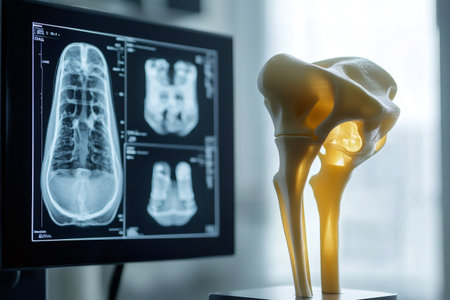
3D printed model of a hip bone and femur standing next to a monitor displaying X ray images, symbolizing medical innovation
Коллекция по умолчанию
Коллекция по умолчанию
Создать новую
Detail of a large fossilized bone
Коллекция по умолчанию
Коллекция по умолчанию
Создать новую
3D illustration of human bones, focusing on a joint, with neutral background and open copy space to the side.
Коллекция по умолчанию
Коллекция по умолчанию
Создать новую
A vibrant skeletal model showcases the structure of human bones and joints, enhancing anatomy learning in a modern science laboratory environment.
Коллекция по умолчанию
Коллекция по умолчанию
Создать новую
Detailed view of a human femur bone against a dark background, highlighting its anatomy and structure.
Коллекция по умолчанию
Коллекция по умолчанию
Создать новую
3d rendered medically accurate illustration of the upper femur joint
Коллекция по умолчанию
Коллекция по умолчанию
Создать новую

X-ray of the hip prosthesis elderly man
Коллекция по умолчанию
Коллекция по умолчанию
Создать новую
Model of a human hip joint displayed during a medical consultation in a clinic setting
Коллекция по умолчанию
Коллекция по умолчанию
Создать новую
A bright, minimalistic setting featuring x-ray displaying a vibrant light blue holographic 3D brain, uses red-orange tones to indicate areas of inflammation or abnormality.
Коллекция по умолчанию
Коллекция по умолчанию
Создать новую
Skeleton, legs and x ray of pelvis on black background for injury assessment, bone diagnostic or
Коллекция по умолчанию
Коллекция по умолчанию
Создать новую
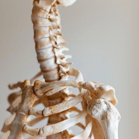
Close-up view of a human skeletal model showcasing the spine and ribcage. Ideal for educational purposes, this image highlights important anatomical details for biology and health studies.
Коллекция по умолчанию
Коллекция по умолчанию
Создать новую
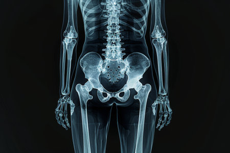
X-ray of human body with bones and joints, medical concept
Коллекция по умолчанию
Коллекция по умолчанию
Создать новую

bones, pelvis, femur, black background, 3d rendering
Коллекция по умолчанию
Коллекция по умолчанию
Создать новую
3d rendered illustration of the hip bone
Коллекция по умолчанию
Коллекция по умолчанию
Создать новую

Two bones rest on a rustic wooden surface surrounded by blurred twinkling lights and greenery capturing the warmth of New Year celebrations.
Коллекция по умолчанию
Коллекция по умолчанию
Создать новую

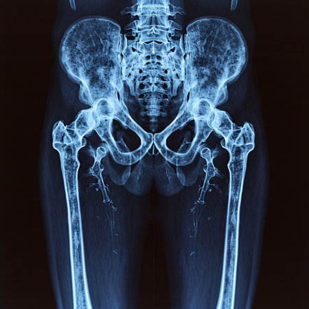
Scanning of an anterior posterior radiograph of the pelvis taken, among others radiographs, to try to detect the origin of pain in the hip of an adult man
Коллекция по умолчанию
Коллекция по умолчанию
Создать новую
Anatomical illustration of the human pelvis showing the sciatic nerve highlighted in pink, indicating pain or inflammation.
Коллекция по умолчанию
Коллекция по умолчанию
Создать новую
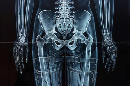
A detailed x-ray revealing the skeletal structure of a human body, focusing on the pelvic area, spine, and limbs, typically used for medical diagnostics.
Коллекция по умолчанию
Коллекция по умолчанию
Создать новую

old bone isolated on white background
Коллекция по умолчанию
Коллекция по умолчанию
Создать новую
A close-up of an X-ray film showing a broken bone, with the fracture clearly visible.
Коллекция по умолчанию
Коллекция по умолчанию
Создать новую

Chicken bones
Коллекция по умолчанию
Коллекция по умолчанию
Создать новую
A close-up view of a broken bone fragment resting on a black background, showcasing the intricate details and shadows, ideal for medical or anatomical illustrations.
Коллекция по умолчанию
Коллекция по умолчанию
Создать новую
A vibrant 3D rendering highlights the human skeleton, focusing on the pelvic area with detailed colors, illustrating anatomical features in a laboratory environment.
Коллекция по умолчанию
Коллекция по умолчанию
Создать новую

photograph of articulated hip joint in black background
Коллекция по умолчанию
Коллекция по умолчанию
Создать новую
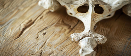
A weathered, white animal bone with a rough surface lies on a wooden table. The bone is partially visible, with the end of the bone pointing towards the bottom right corner of the frame. The wooden surface appears to be old, with visible grain and imperfections.
Коллекция по умолчанию
Коллекция по умолчанию
Создать новую

X ray of Hip joint pain bone
Коллекция по умолчанию
Коллекция по умолчанию
Создать новую
x-ray of a human hip , black background
Коллекция по умолчанию
Коллекция по умолчанию
Создать новую
Doctor man pointing on arm of human skeleton anatomical model. Physiotherapist explaining joints model. Chiropractor or osteopath points to the skeleton of human body. Bones anatomy close up
Коллекция по умолчанию
Коллекция по умолчанию
Создать новую
X ray film of pelvic bone fracture
Коллекция по умолчанию
Коллекция по умолчанию
Создать новую
medically accurate 3d illustration of the painful hip
Коллекция по умолчанию
Коллекция по умолчанию
Создать новую
Female doctor analyzing holographic 3d pelvis x ray scan with blurred background
Коллекция по умолчанию
Коллекция по умолчанию
Создать новую
X-ray revealing sharp edges of broken ceramic piece showcasing fragility and fragmentation
Коллекция по умолчанию
Коллекция по умолчанию
Создать новую

doctor's palm with a holographic graphic Bone X-ray generative ai
Коллекция по умолчанию
Коллекция по умолчанию
Создать новую
Anatomical model of human pelvis standing on doctors desk in medical office
Коллекция по умолчанию
Коллекция по умолчанию
Создать новую
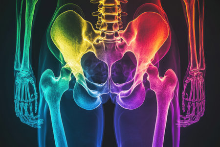
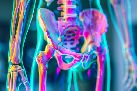
Vivid anatomical representation of the human pelvis and lower limbs, displaying the skeletal structure in a spectrum of colors. This detailed view highlights bone connections and anatomy.
Коллекция по умолчанию
Коллекция по умолчанию
Создать новую
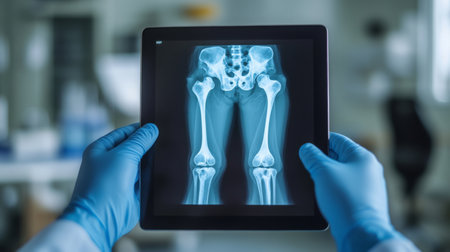
Medical professional examines x-ray of human leg bones using advanced tablet technology
Коллекция по умолчанию
Коллекция по умолчанию
Создать новую
X-ray image of the pelvis and hip of a patient
Коллекция по умолчанию
Коллекция по умолчанию
Создать новую
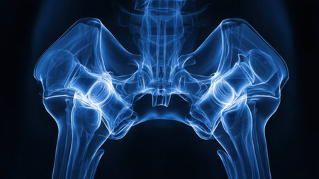
High-resolution X-ray image showcasing the intricate details of human hip joint anatomy, focusing on the pelvis and its components for educational use.
Коллекция по умолчанию
Коллекция по умолчанию
Создать новую
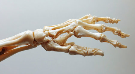
A close-up representation of a human hand skeleton highlights the intricacies of the phalanges and metacarpals
Коллекция по умолчанию
Коллекция по умолчанию
Создать новую

3d rendered illustration of an inflamed coccyx
Коллекция по умолчанию
Коллекция по умолчанию
Создать новую
An x-ray image showcasing the intricate details of a human skeleton, Human hip bones showing in X-ray film, AI Generated
Коллекция по умолчанию
Коллекция по умолчанию
Создать новую
X-ray of human foot. X-ray of human foot. Vector illustration
Коллекция по умолчанию
Коллекция по умолчанию
Создать новую
Legion-Media
Создайте свои проекты на основе качественных стоковых фотографий и видео.


Copyright © Legion-Media.